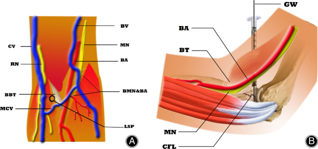
Fig. 2.

Schematic orientation of the syringe guide placement. Front view (A): BA, brachial artery; BBT, biceps brachii tendon; BMN & BA, branches of the median nerve and brachial artery; BV, basilic vein; CV, cephalic vein; LSP, location of syringe placement; MCV, median cubital vein; MN, median nerve; RN, radial nerve. Medial view (B): BA, brachial artery; BT, biceps tendon; CFL, coronoid fracture line; GW, guide wire; MN, median nerve.
