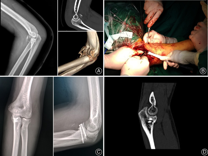
Fig. 4.

Intra‐op screw placement in a comminuted coronoid fracture. Lateral X‐ray and CT scan (sagittal view) of a comminuted coronoid fracture (A), intra‐operative screws fixation of the coronoid fracture (B), post‐operative day seven AP and lateral X‐rays of the coronoid process (C), CT scan (sagittal view) of the coronoid process 1‐month post‐op (D).
