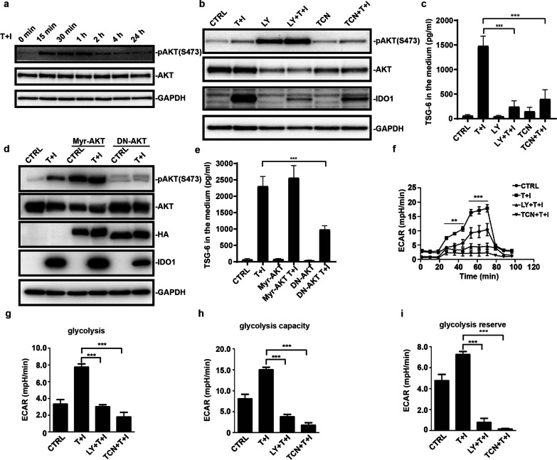
Fig. 3.
TNFα and IFNγ acutely increase the phosphorylation of AKT. a MSCs were stimulated with TNFα and IFNγ (10 ng/ml each) for different periods and lysed. Total AKT and phosphorylated AKT (S473) were examined by western blotting. b MSCs were pretreated with LY294002 (LY) or triciribine (TCN) for 1 h and then TNFα and IFNγ were added into the medium for 24 h. MSCs were lysed and total protein was extracted. IDO1, total AKT, and phosphorylated AKT (S473) were examined by western blotting. c MSCs treated as in (b), and TSG-6 in the medium were examined by ELISA. d MSCs were infected by constitutive active form Myr-AKT or dominant-negative AKT (DN-AKT) adenovirus two days before TNFα and IFNγ stimulation. After TNFα and IFNγ treatment for 24 h, MSCs were lysed and total protein was extracted. IDO1, total AKT, and phosphorylated AKT (S473) were examined by western blotting. HA-tagged Myr-AKT or HA-tagged DN-AKT was immunoblotted by antibody against HA. e MSCs treated as in (d), and TSG-6 in the medium were examined by ELISA. f Extracellular acidification rate (ECAR) of hUC-MSCs was monitored when TNFα and IFNγ treatment for 1 h. g The basal and (h) maximal glycolysis values in figure (f) were statistically analyzed. (i) Calculation of the glycolysis reserve in figure (f) is subtraction basal glycolysis from maximal glycolysis. Data are presented as mean ± SEM. of triplicates (d–i) ***, p < 0.001

