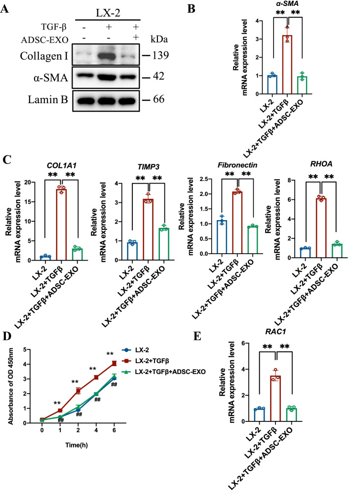
Fig. 4.
ADSC-EXO treatment suppresses HSCs activation and attenuates its profibrogenic phenotype in vitro. A Western blot analysis of HSCs markers α-SMA and Collagen I protein level in untreated LX-2 cells, LX-2 cells with 12 h TGF-β treatment (10 ng/mL) for activation and activated LX-2 cells exposed to ADSC-EXO (200 μg/ml) for 24 h. GAPDH as internal reference. B Relative mRNA expression level of α-SMA in LX-2 cells with different treatments was determined by qRT-PCR analysis (3 replicates). C Relative mRNA expression level of profibrogenic-associated genes COL1A1, TIMP3, Fibronectin, RHOA in LX-2 cells was determined by qRT-PCR analysis (3 replicates). D CCK8 assay was performed for LX-2 cells proliferation ability measurement in each group. 1 h, 2 h, 4 h and 6 h represent ADSC-EXO exposure duration (3 replicates). E Relative mRNA expression level of cell proliferation marker RAC1 in LX-2 cells was determined by qRT-PCR analysis (3 replicates). All data are shown as the mean ± SEM, *p < 0.05, **p < 0.01 comparing with corresponding controls by unpaired t test between two groups

