
Figure 4: Treatment with DOTA-EB-TATE promotes tumor regression and progression-free survival in high-SSTR2-expressing tumors.
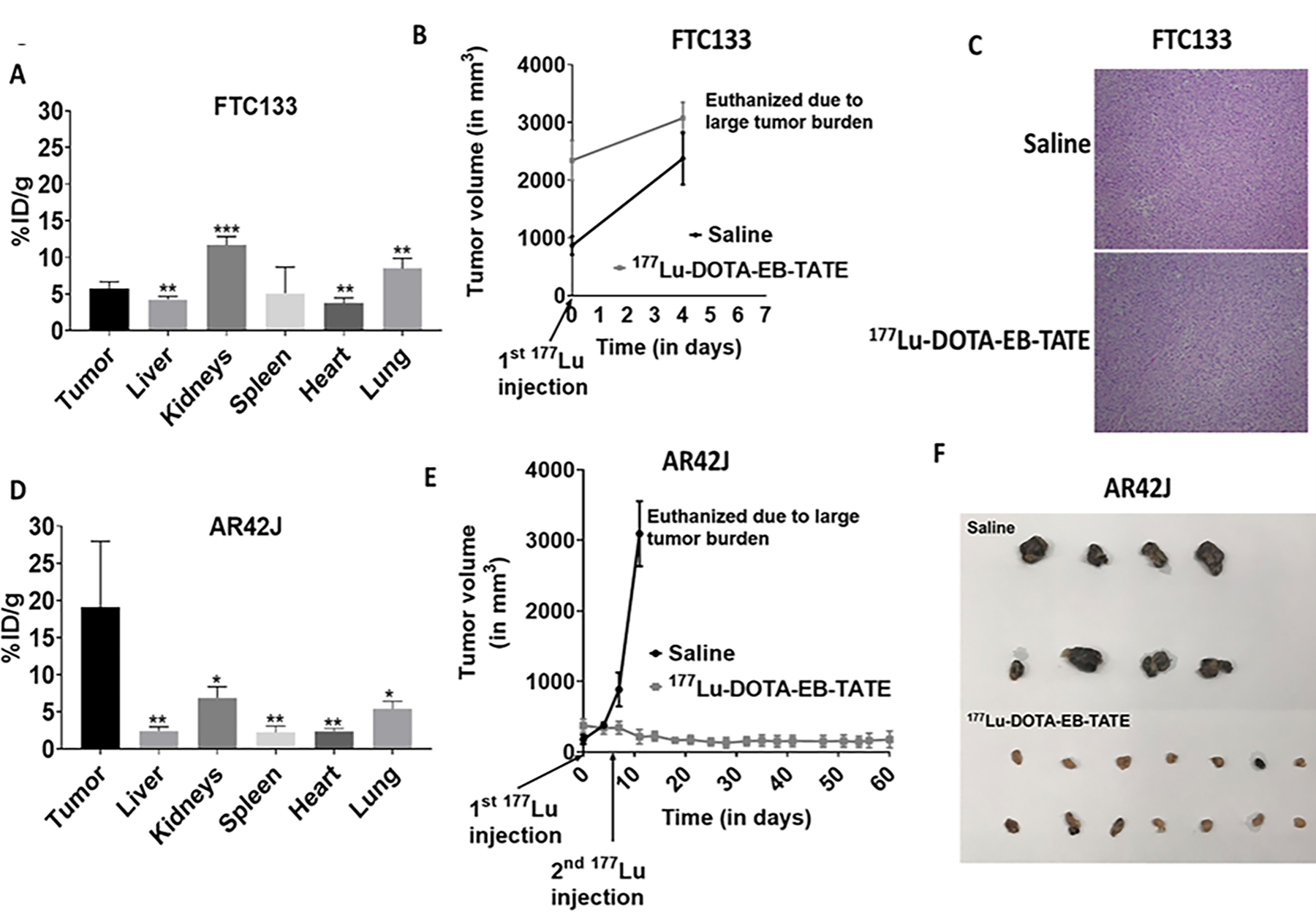
(A) The bar graph shows quantification of 86Y-DOTA-EB-TATE in the tumor and normal tissues (liver, kidneys, spleen, heart, and lungs) of the FTC133 subcutaneous xenograft mice (n=5). ***p<0.001, **p<0.01 w.r.t tumor tissue.
(B) No effect of PRRT on FTC133 (low-SSTR2-expressing) subcutaneous xenograft mice receiving 177Lu-DOTA-EB-TATE treatment (n=8) in comparison to the mice that received saline (n=4). Data are presented as mean±SEM.
(C) Representative hematoxylin-eosine (H-E) stained slides show no difference in the histology of FTC133 tumors derived from the saline and 177Lu-DOTA-EB-TATE treated mice.
(D) The bar graph shows quantification of 86Y-DOTA-EB-TATE in the tumor and normal tissues (liver, kidneys, spleen, heart, and lungs) of the AR42J subcutaneous xenograft mice (n=4). **p<0.01, *p<0.05 w.r.t tumor tissue.
(E) Significant reduction in the tumor volume of the AR42J (high-SSTR2-expressing) subcutaneous xenograft mice that received 177Lu-DOTA-EB-TATE treatment (n=8) in comparison to the saline treated mice (n=4). Data are presented as mean±SEM.
(F) Representative images of the tumor tissues collected after euthanasia from saline and 177Lu-DOTA-EB-TATE treated AR42J subcutaneous xenograft mice.