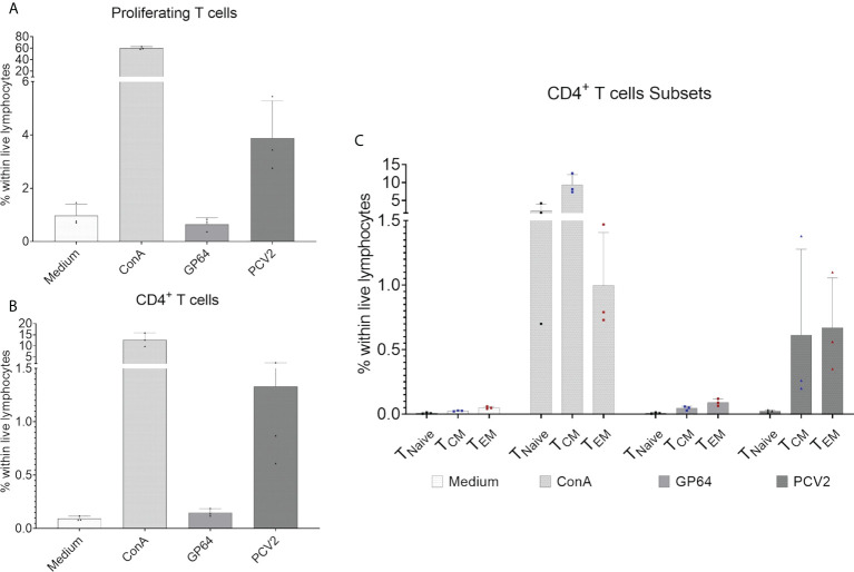
Figure 8.
Proliferation assay of CellTrace™Violet-stained PBMCs after restimulation with baculovirus-expressed PCV2-ORF2 protein. PBMCs cultivated in medium or stimulated with an irrelevant baculovirus-expressed protein (GP64) were used as negative controls, ConA-stimulated PBMCs served as positive control. (A) Shows the percentage of reacting T cells within live lymphocytes of three representative EGMs. (B) shows the percentage of proliferating CD4+ T cells within live lymphocytes of three EGMs. (C) Shows the reactivity of the CD8α/CD27-defined CD4+ T- cell subpopulation of three EGMs. Naive CD4+ T cells (TNaive), central memory CD4+ T cells (TCM) and effector memory CD4+ T cells (TEM).

