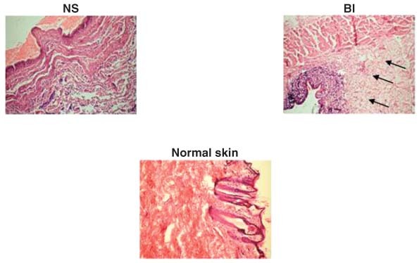
Figure 1.

Photomicrographs showing healing pattern on day 3 wounds treated with normal saline (NS), and autologous bone marrow‐derived cells injected to wound margins (BI). Healing tissue in BI‐treated wound was better with compact and wider granulation tissue (arrows) than NS‐treated wound (H&E sections 10×, day 3).
