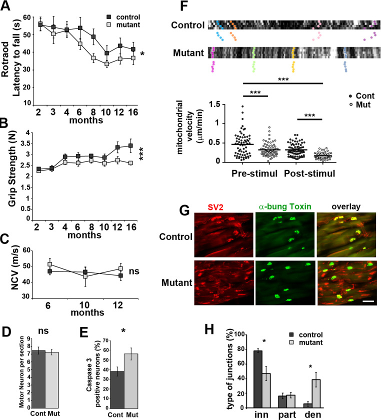
Fig 3. Mutant mice show motor deficit and display axonal defects.
A- Rotarod latency to fall and B- grip strength were measured between 2 and 16 months postnatal on Control (n = 21) and Mutant (n = 25) mice. Two-way non repeated measures ANOVA Sidak’s post-hoc tests. C- Nerve conduction velocity (NCV) measured in Control and Mutant mice (n = 10 each). Two-way non repeated measures ANOVA Sidak’s post-hoc tests. D- Spinal cord sections of Mutant (Mut) and Control (Cont) mice (12 months) were stained with cresyl violet and motor neurons were counted (see S8 Fig in S1 File). n = 3 animals (14 to 30 sections). Two tailed Student t-test. E- Spinal cord sections of Mutant (Mut, n = 19 images, 3 animals) and Control (Cont, n = 12 images, 2 animals) mice were immunostained for Caspase 3, ChAT and Neurofilament (S8 Fig in S1 File) (12 months old). Caspase 3 positive neurons in percentage of Neurofilament positive neurons. Two tailed Student t-test. F- Axonal mitochondria labelled with mito-Dsred2 were imaged in vivo before and after electrical stimulation. Upper panels show typical kymographs. Tracked mitochondria are shown with colour dots. Mutant mice mitochondria follow a straight pattern indicating they are immobile or slowly moving. Lower panel: Mitochondria speed was plotted according to the genotype before and after stimulation. One-way ANOVA Tukey’s post-hoc test. n are provided in Material and Methods. G- Gastrocnemius muscle neuromuscular junctions of 12 months old Mutant and Control mice were stained for presynaptic SV2 and postsynaptic acetylcholine receptor with FITC-α-bungarotoxin (α-bung). Scale bar = 100μm. H- Innervated (inn, complete overlap), partially innervated (part, partial overlap) and denervated (den, no overlap) junctions were counted on sections as shown in D. Two tailed Student t-test. n = 4 mice (12 months). Error bars represent SEM.

