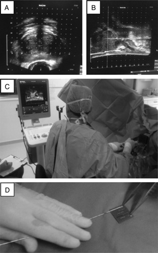
FIGURE 1.

Transperitoneal template biopsy in practice. A, Initial assessment of possible core biopsy sites is made using the coronal view of a transrectal ultrasound scan of the prostate. B, Sagittal transrectal ultrasound scan view of the prostate during core biopsy sampling; short arrow indicate the cranial limit of the prostate; long arrow indicates the needle within the prostate. C, Core biopsy needle is inserted using the template as a guide and observed in real time under transrectal ultrasound scan; note assistant in left bottom corner annotating biopsy sites on proforma. D, Blotting of the core of tissue from the needle onto the sponge and (inset) 6 consecutive cores labeled and ready to be overlaid with a wet sponge.
