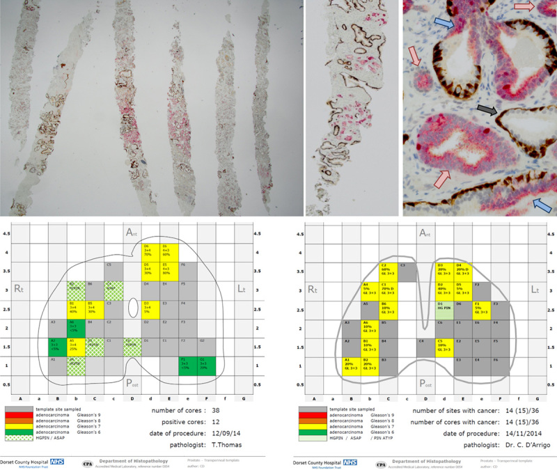
FIGURE 2.

Histopathology report on the 2-dimensional map and multiplex immunohistochemistry. A–C, Multiplex immunohistochemistry at x40, x100 & x400. CK5 and p63 (basal cell markers) stained in brown, racemase, and Ki-67 stained in red. A (×1.25), Substantial amount of invasive carcinoma can be seen in all cores, characterized by the absence of brown staining; some of the carcinoma expresses racemase (red) other is racemase(−). B (×2), Scattered clusters of invasive carcinoma devoid of brown staining and racemase(+) are seen clearly among the background of normal prostatic glands that have brown staining but lack racemase. C (×10), High-grade prostatic intraepithelial neoplasia (blue arrows) have discontinuous basal cells (brown staining) and can be readily distinguished from acini of invasive carcinoma (red arrows) and atrophic glands (gray arrow). D and E, Examples of histopathology reports with 2-dimensional maps using the visual traffic light coded system.
