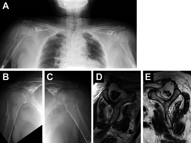
Figure 4. Case 2. Preoperative radiographic images.
The plain radiographs (A-C) show bilateral PHFs. The oblique sagittal T2-weighted images on MRI (D: right, E: left) represent muscle atrophy with/without fatty infiltration at the upper part of the subscapularis, supraspinatus, and infraspinatus muscles at bilateral shoulders.

