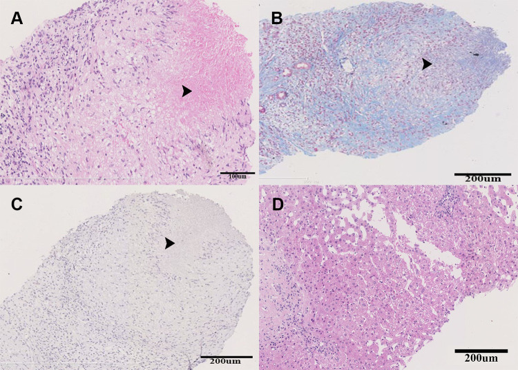
Figure 2.
The histopathology of the liver specimen obtained from the biggest nodule. The black arrows indicated the granulomatous inflammation with central caseous necrosis. (A) Hematoxylin & eosin staining, (B) Masson triple staining, (C) acid-fast staining, and (D) Hematoxylin & eosin staining of the para-nodule liver specimen.

