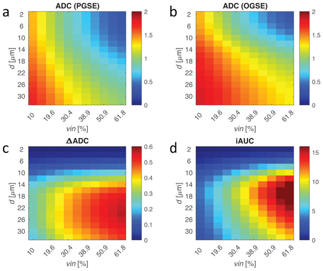
Figure 2.

Finite difference simulations of regularly packed cells with varying cell diameter (d) and volume fraction (vin) using 70 ms (PGSE) and 10 ms (OGSE) diffusion times. (a-b) ADC is fitted from signals with both diffusion times. The diffusion time dependence using (c) the difference in ADC and (d) the iAUC change the sensitivity landscape. Where ADC is mostly sensitive to volume fraction at small restriction sizes, such as axons (1-5 μm) or glia (5-10 μm), ΔADC and iAUC filter out these components to give contrast driven by size-selective cellular density. iAUC provides more selective sensitivity to cancer cells ~10-20 μm than ΔADC.
