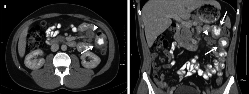
Fig. 1.
Case report: oral and intravenous portal phase contrast-enhanced CT axial (a) and coronal (b) of a 33-year-old male patient diagnosed with left lower eyelid melanoma which subsequently metastasised to the lymph nodes, lungs, brain, SB and subcutaneous tissue in the following 5-year period. Complete response was achieved after chemotherapy, immunotherapy and radiotherapy. One year after treatment completion he relapsed, presenting with two jejunal metastases (arrows) and involvement of the draining mesenteric lymph nodes (arrow heads). Surgical resection was then performed with no evidence of disease recurrence to date, after 10 years of follow-up

