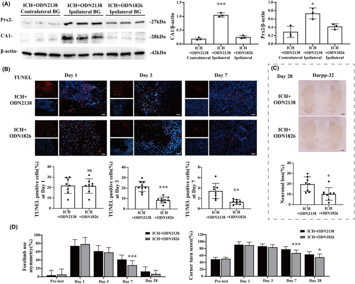
FIGURE 3.

TLR9 activation alleviated neural injury and facilitated functional recovery after ICH. (A) Western blot analysis of CA1 and Prx2 protein levels in ipsilateral BG of ODN2138‐treated mice and ODN1826‐treated mice at day 3 after ICH. n = 3 for each group. *p < 0.05, ***p < 0.001 vs. other groups by one‐way ANOVA. (B) TUNEL staining (red) with DAPI (blue) of ODN2138‐treated mice and ODN1826‐treated mice ipsilateral BG at day 1, day 3, and day 7 after ICH, and the TUNEL‐positive cell percentages of each groups were quantified. n = 8 for each group. Scale bar = 100 μm. **p < 0.01, ***p < 0.001 vs. ICH + ODN2138 group by Student's t‐test. (C) Representative DARPP‐32 immunoreactivity of ODN2138‐treated mice and ODN1826‐treated mice at day 28 after ICH. n = 8 for each group. Scale bar = 500 μm. *p < 0.05 vs. ICH + ODN2138 group by Student's t‐test. (D) Forelimb use asymmetry and corner turn scores before and after ICH with ODN2138 or ODN1826 treatment. n = 44 for each group at day 1 and pretest, n = 33 for each group at day 3, n = 19 for each group at day 7, and n = 8 for each group at day 28. Values are mean ± SD. *p < 0.05, ***p < 0.001 vs. ICH + ODN2138 group by two‐way ANOVA
