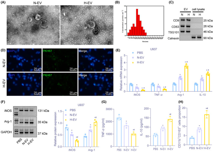
FIGURE 1.

Hypoxic glioma cell‐derived EVs promote M2 macrophage polarization. (A) Images of EVs observed under a TEM (Scale bar = 100 nm). (B) Diameter of EVs detected by dynamic light scattering. (C) The expression of CD9, CD63, TSG101, and calnexin on the surface of EVs determined by Immunoblotting. (D) Detection of the uptake of EVs from normoxic and hypoxic glioma cells by macrophages U937 through immunofluorescence (400×). (E) The mRNA expression of iNOS, TNF‐α, Arg‐1, and IL‐10 assessed by RT‐qPCR after EV treatment. (F) Immunoblotting of protein expression of iNOS and Arg‐1 in macrophages U937 after EV treatment. (G) The expression of TNF‐α and IL‐10 in supernatant of macrophages U937 evaluated by ELISA assay. (H) The proportion of CD11b+CD163+ cells in U937 cells examined by flow cytometry. *p < 0.05 versus macrophages treated with PBS. # p < 0.05 versus macrophages treated with normal oxygen‐induced glioma cell‐derived EVs. The experiment was repeated three times
