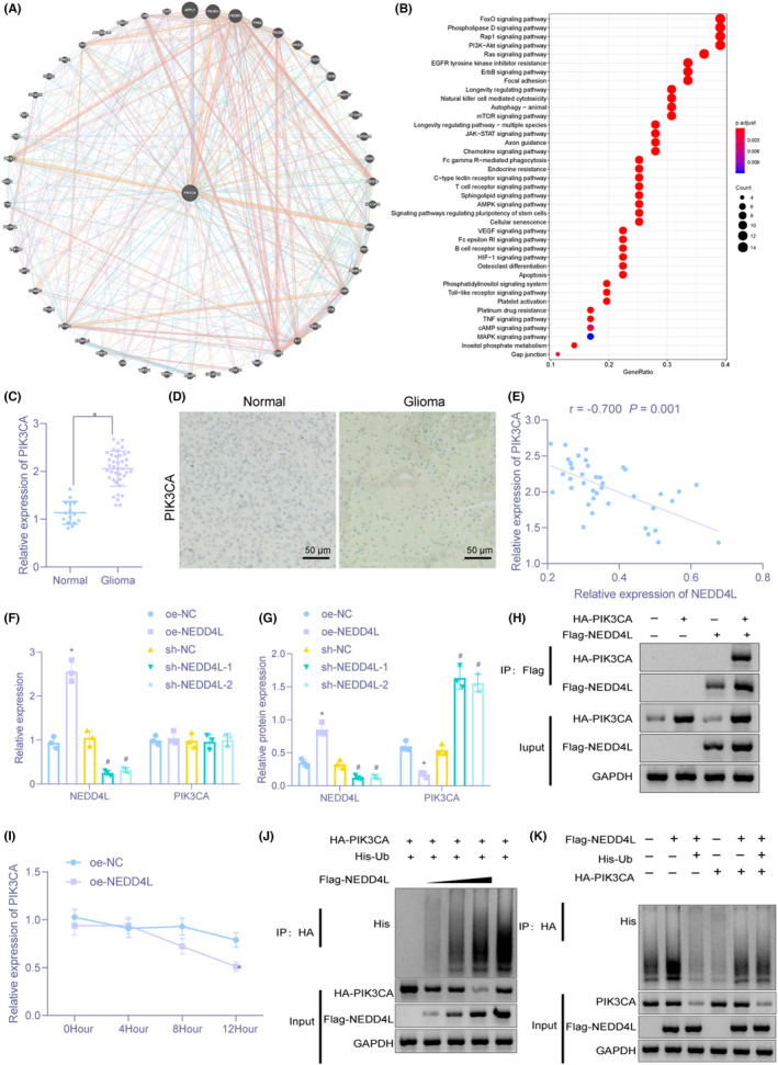
FIGURE 5.

NEDD4L enhances ubiquitination and degradation of PIK3CA in macrophages. (A) PIK3CA‐related gene prediction. Circle center denotes PIK3CA gene, and others are predicted related genes. (B) KEGG pathway enrichment analysis of PIK3CA‐related genes. The x‐axis represents GeneRatio, the y‐axis represents the KEGG entry name, and the histogram on the right is color scale. (C) The expression of PIK3CA in brain tissues of 40 cases of glioma and 15 cases of non‐glioma examined by RT‐qPCR. (D) The expression of PIK3CA in brain tissues of 40 cases of glioma and 15 cases of non‐glioma assessed by immunohistochemistry (×200). (E) NEDD4L expression negatively correlated with PIK3CA expression identified by Pearson correlation analysis. (F) The mRNA expression of NEDD4L and PIK3CA in U937 cells evaluated by RT‐qPCR. (G) Immunoblotting for measurement of protein expression of NEDD4L and PIK3CA in U937 cells. (H) Interaction between NEDD4L and PIK3CA verified by Co‐IP assay. (I) The protein expression of NEDD4L and PIK3CA in U937 cells with 20–40 μg/ml CHX treatment determined by Immunoblotting. (J) The effect of NEDD4L on PIK3CA ubiquitination in HEK293T cells detected by IP assay. (K) The impact of NEDD4L on endogenous PIK3CA ubiquitination in HEK293T cells examined by IP assay. *p < 0.05 versus brain tissues from patients with non‐glioma or U937 cells transfected with oe‐NC. # p < 0.05 versus U937 cells transfected with sh‐NC. The experiment was repeated three times
