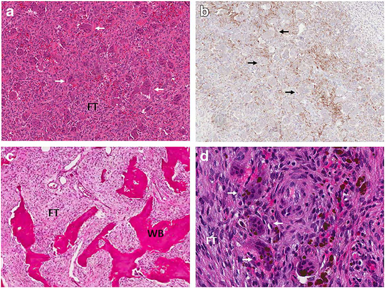
Fig. 1.
Representative histologic images. Upper panels show sections from a giant cell tumor. a Hematoxylin and eosin staining shows characteristic areas of fibrous tissue (FT) interspersed with osteoclast-like giant cells (white arrows). b RANKL immunostaining shows positivity in neoplastic stromal cells. Prominent giant cells are again visualized (black arrows). Lower panels show sections from a fibrous dysplasia lesion. c Hematoxylin and eosin staining shows characteristic areas of fibrous tissue (FT) interspersed with discontinuous trabeculae of abnormal woven bone (WB). d High-power view shows giant cells (white arrows) amidst a background of fibrous tissue (FT) comprised of neoplastic stromal cells

