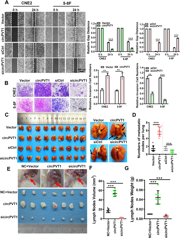
Fig. 2.
circPVT1 promotes the migration and invasion of NPC cells in vitro and NPC metastasis in vivo. A. Wound healing assay showed that circPVT1 promoted CNE2 and 5-8F cell migration. Images were acquired at 0 and 24 h. Data were represented as mean ± SD. ***, p < 0.001. B. Transwell assay showed that circPVT1 promoted CNE2 and 5-8F cell invasion. Data were represented as mean ± SD. **, p < 0.05; ***, p < 0.001. C. Images of visible nodules on the lung surface. CNE2 cells transfected with empty vector, circPVT1 overexpression vector, scrambled siRNA, or sicircPVT1 were injected into nude mouse tail vein (n = 9 for each group), and mice were sacrificed 8 weeks later. Arrows showed visible nodules on the lung surface. D. Quantification of lung metastatic nodules on lung surface. Data were represented as mean ± SD (n = 9 per group). ***, p < 0.001. E. Representative images of mice lymph nodes in the popliteal fossa of mice after injection with transfected CNE2 cells. F-G. Lymph nodes volumes (F) and lymph nodes weights (G) were measured for each group (n = 7 per group). Data were represented as mean ± SD. ***, p < 0.001

