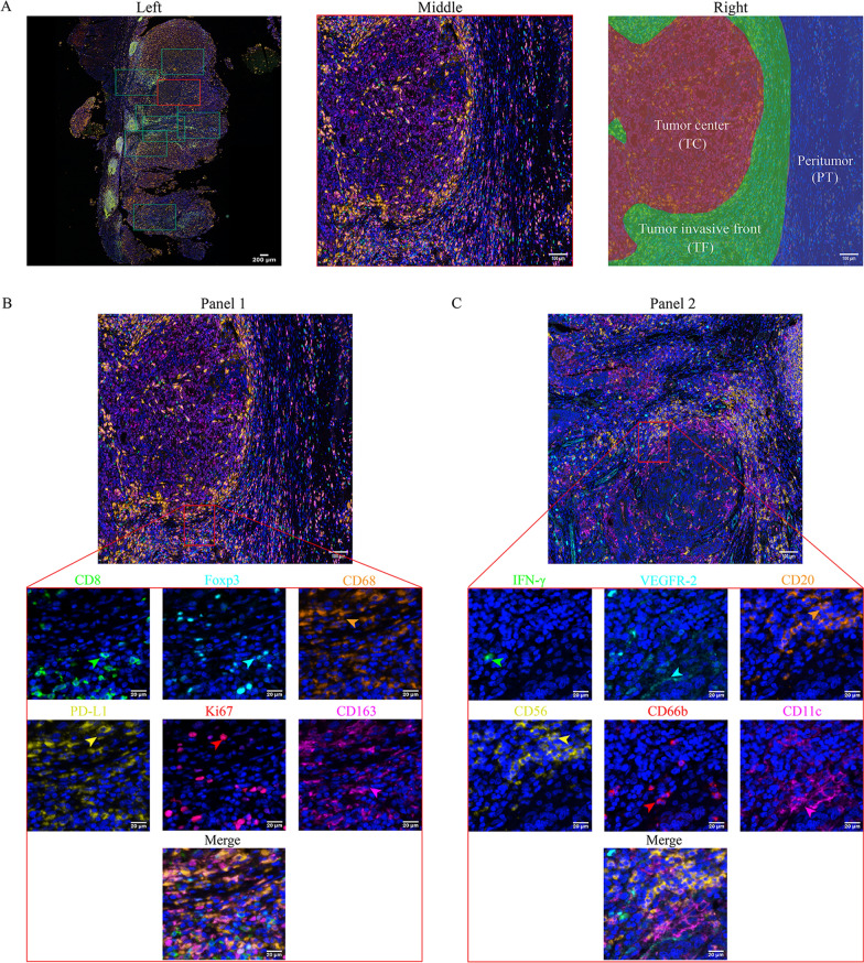
Fig. 1.
Image acquisition, partitioning and staining scheme. A left side: 10× scan of the whole film; middle side: 20× local area magnification imaging; right side: tumor center, tumor invasive front (less than 150 µm from the tumor) and peritumor (more than 150 µm from the tumor); B Staining Panel l (the green, cyan, orange, yellow, red and magenta arrows indicate positive cells with the expression of CD8, Foxp3, CD68, PD-L1, Ki67 and CD163 proteins in tumor tissue); C Staining Panel 2 (the green, cyan, orange, yellow, red and magenta arrows indicate positive cells with the expression of INF-γ, VEGFR-2, CD20, CD56,CD66b and CD11c proteins in tumor tissue)

