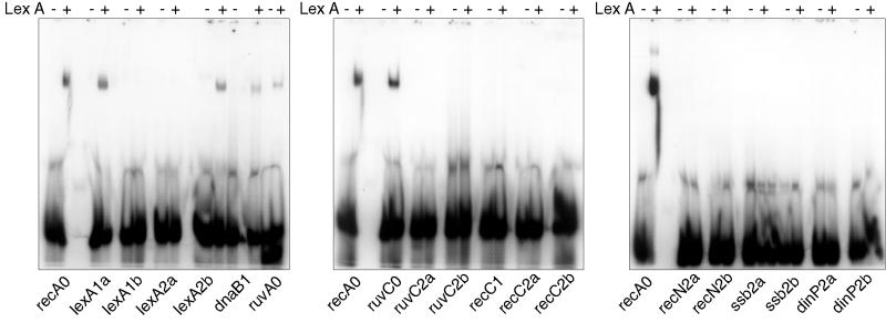
FIG. 3.
Analysis of LexA binding to potential SOS boxes. Double-stranded oligonucleotides containing each identified motif (indicated below the gel) were end labeled with [γ-32P]dATP; following incubation with 8 nM (final concentration) purified M. tuberculosis LexA (lanes marked +) they were assessed for LexA binding by gel shift compared with no-protein controls (lanes marked −). The recA0 SOS box, which had been shown previously to bind LexA, was included on each gel as a positive control. The figure was compiled using Adobe Photoshop.

