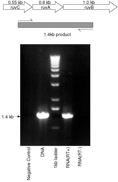
FIG. 5.
The ruvCAB genes are cotranscribed. The top part shows schematically the arrangement of the ruv genes in M. tuberculosis, with the positions of the primers used for RT-PCR and the size of the expected product indicated. The lower part shows that the product obtained using RNA which has been reverse transcribed is the same size as that obtained by PCR using chromosomal DNA, while no product is formed using RNA if the RT step is omitted. The figure was compiled using Adobe Photoshop and Macromedia Freehand.

