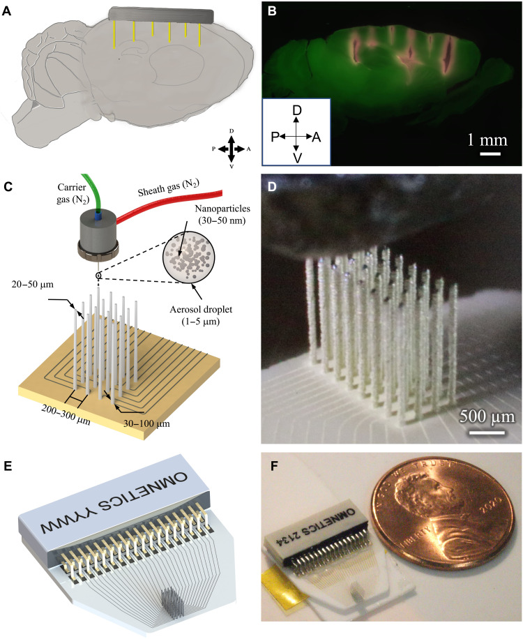
Fig. 1. Example use-case and printing process.
(A) To understand circuit interactions, the activity of multiple brain areas must be recorded. The target brain areas will differ for any given study. In this conceptual schematic, the target areas to hit are (from left to right) mouse prefrontal cortex, motor cortex, caudal striatum, and hippocampal CA2 and CA3. (B) Slice of an actual mouse brain demonstrating the targeting of the brain areas mentioned in (A). The sagittal slice shows penetration of several 3D printed shanks (red) of a single probe into cortex, striatum, and hippocampus. Penetrations were parallel; appearance of shank trajectories are due to tissue dehydration in histological processing. (C) Schematic of AJ 3D printing of metal nanoparticles rapidly creates structurally robust, fully customizable neural probes, including the circuit board for routing of physiological signals. (D) Image during printing of a 32-channel probe. Also see movies S1, S2, and S3 for the printing process. (E) Schematic design of a 32-channel probe with routing and attached connector. (F) Probe printed on the basis of the design shown in (E) with a U.S. one cent coin for scale.

