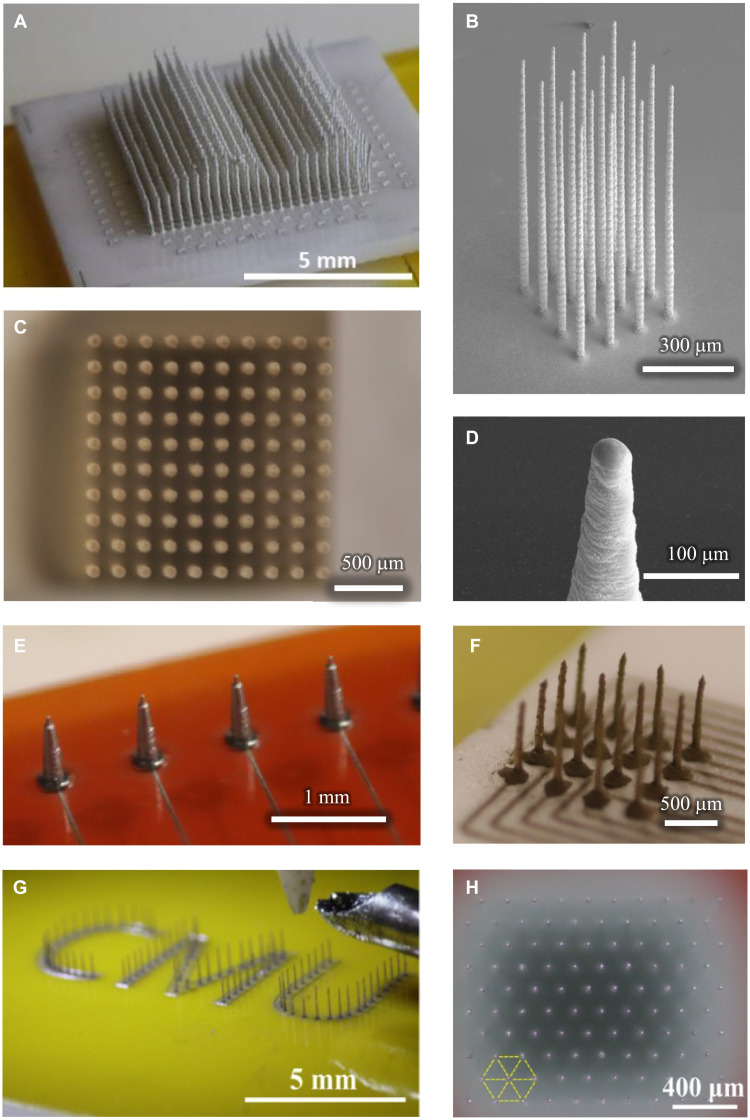
Fig. 2. CMU array fabrication using 3D nanoparticle printing.
(A) A 512-electrode shank array with variable shank heights demonstrating the customization possible by this method. Shank lengths are stepped to demonstrate regularity; individual shank lengths can be designated independently and to any value. A time-lapse video of the construction of this probe is shown in movie S2. (B) Printed platform with 1-mm-long shanks having an aspect ratio of 1:50. The substrate is alumina ceramic. It only takes under 1.5 min to create a 1-mm-long shank with this method. (C) A printed 10 × 10 array with a height of 2.4 mm and average width of 30 μm reveals straight and uniform shanks when viewed top-down. A similar geometry is used through the manuscript. (D) Close-up image of tapered shank tip, which facilitates their insertion into tissue. (E) Example of shanks directly printed on a flexible polyimide (Kapton) substrate. Printed wiring on the same substrate can enable connection to data acquisition hardware. (F) Example of shanks printed with gold nanoparticles, here demonstrating a wider base for increased robustness. Note that to demonstrate the diversity of geometries, the other panels in this figure are printed with less expensive silver ink. (G) Shank customization in any arrangement is possible: Here, shanks are printed to resemble the Carnegie Mellon University abbreviation “CMU.” (H) High-density, high–aspect ratio array printed in hexagonally packed pattern with 200-μm pitch (view from above). A 15% improvement in areal electrode density can be achieved by pattering the electrodes in hexagonal pattern rather than square packing.

