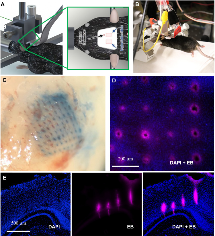
Fig. 6. Probe insertion into mouse brain.
(A and B) Schematic and photograph of form factor used during insertion for recording using a standard stereotaxic micromanipulator. (C) Evans blue dye left behind from the successful insertion of a dense (2600 shanks/cm2; pitch of 200 μm), 10 × 10 array of 20-μm tip diameter. (D) No gross tissue damage was observed following 30 min of insertion [blue, 4′,6-diamidino-2-phenylindole (DAPI) nuclear stain; red, Evans blue (EB)]. (E) Successful insertion test in an anesthetized mouse without breaking the shanks or gross damage to the brain (coronal slice though area V2, hippocampus). Note that the lack of damage or tearing is caused by probe insertion in (C) to (E).

