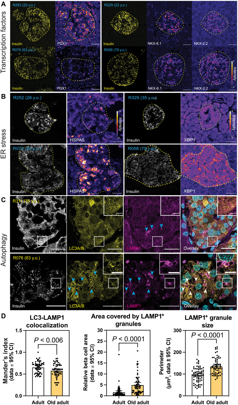
Fig. 2. Beta cell TFs, ER stress, and autophagy markers in aging beta cells.
Immunohistochemistry and confocal microscopy of human pancreas formalin-fixed paraffin-embedded samples from adults (22 to 35 years old) and old adults (61 to 79 years old). Human islets were stained with (A) insulin, PDX1, NKX6-1, and/or NKX2-2; (B) insulin and HSPA5 or XBP1; and (C) insulin, LC3A/B, and LAMP1. (D) Quantification of LC3-LAMP1 colocalization, area of beta cell cytosol covered by LAMP1+ granules, and LAMP1+ granule size. Each dot represents data from a single region of ~140 μm2 of human islets. Here, 1 region per islet, 10 regions per donor were quantified. In (A) and (B), the islet region is demarcated by the dotted yellow line. Scale bars, 50 μm (A and B), 20 μm (C), and 5 μm (inset of C). CI, confidence interval.

