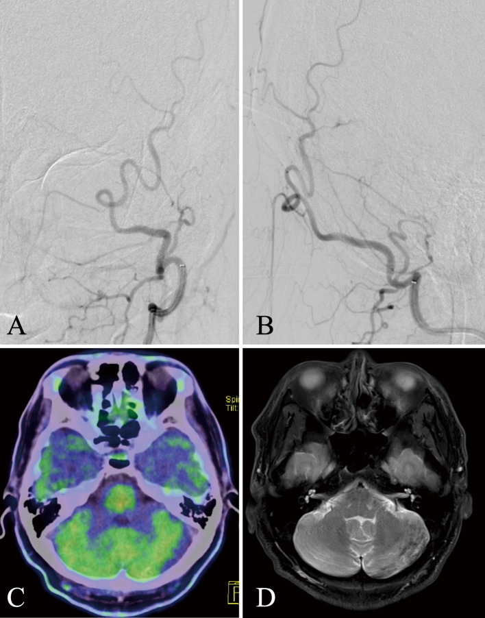
Fig. 3.
Postoperative images of digital subtraction angiography, positron emission tomography, and T2-weighted image.
A and B: Frontal and lateral view of left-sided occipital angiography after Onyx transarterial embolization; the shunt flow disappeared completely. C: Six months after surgery, the high preoperative uptake found in methionine-positron emission tomography had disappeared. D: T2-weighted image. The abnormal hyperintense area in the left cerebellum had also been extensively improved.

