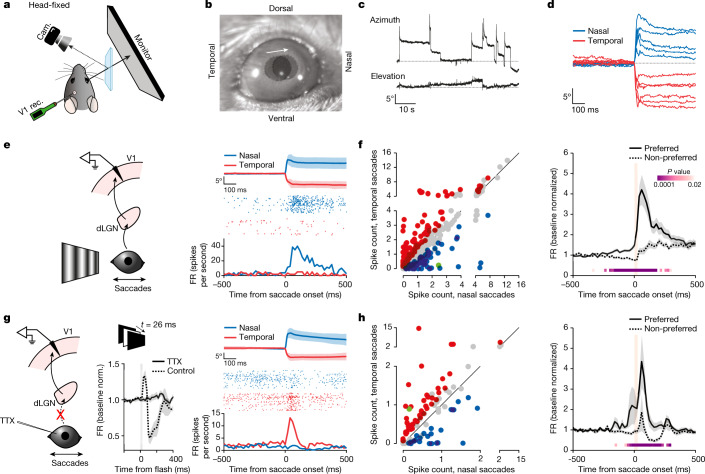
Fig. 2. Direction-selective non-visual V1 response to saccades.
a, Experimental set-up in head-fixed mice. b, Two overlaid snapshots (before and after a nasal saccade). The arrow indicates saccade direction. Circles delineate the pupils. c, Example eye position traces. Top, azimuth (up is nasal); bottom, elevation (up is dorsal). d, Example azimuthal eye position for nasal and temporal saccades. e, Left, schematic of V1 recording during saccades on a vertical grating. Right, example neuron. Average eye position for nasal and temporal saccades (top; shaded area, average ± s.d.), raster plots (centre) and the PETH (bottom) are shown. f, Left, scatterplot of the response to nasal and temporal saccades (average spike count in a 100-ms window from saccade onset), for all responsive neurons (n = 415 neurons, 13 mice). Blue, nasal preference; red, temporal preference; grey, no statistical difference; green, example in e. Right, average PETH of discriminating neurons (n = 192 neurons, 13 mice), for preferred and non-preferred directions. Shaded area, average ± s.e.m. Orange bar, 0–90% rise time of saccades (26 ms). P values are from the comparison of activity for preferred and non-preferred saccade directions in 20-ms bins (Wilcoxon signed-rank test, one tailed). g, Left, schematic of V1 recording during saccades in TTX-blinded animals. Centre, average multi-unit responses to a brief full-field flash. Grey bar, flash duration (26 ms). Note the lack of response with TTX. Control, 137.9 ± 31.2 Hz (FR ± s.e.m. averaged over a 60-ms window 10 ms after response onset; n = 4 mice; 22.1% ± 4.3% increase over baseline); TTX, −8.0 ± 15.4 Hz (0.12% ± 3.0% increase over baseline; n = 8 mice). Wilcoxon rank-sum test, one tailed: P = 0.0020. Right, example neuron in a TTX-blinded animal. Average eye position (top; shaded area, average ± s.d.), raster plots (centre) and the PETH (bottom) are shown. h, Same as in f, but for TTX-blinded animals. Left, n = 97; right, n = 67 (8 mice). Green, example neuron in g. 0–90% rise time of saccades, 27 ms.

