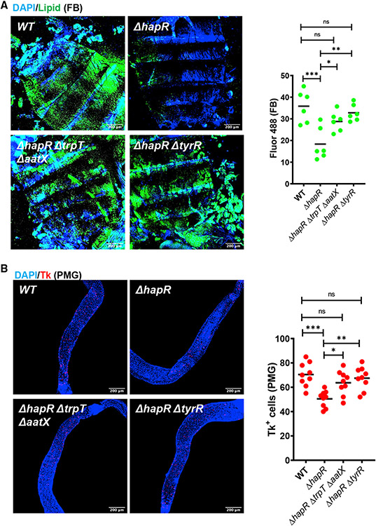
Figure 5. Decreased tryptophan uptake by the V. cholerae ΔhapR mutant preserves host fat stores and increases Tk expression in the PMG during infection.
(A) Representative micrographs and fluorescence quantification (lipid) in fat bodies of OreR flies infected with the indicated V. cholerae strains and stained with Bodipy and DAPI.
(B) Representative micrographs of Tk immunofluorescence in the PMG of flies analyzed in (A). Flies were dissected after 48 h of infection with the indicated V. cholerae strain. Scale bars, 200 μm. At least six intestines were evaluated. The mean is shown. A one-way ANOVA with Tukey’s multiple comparisons test was used to calculate statistical significance. ***p < 0.001, **p < 0.01, *p < 0.05.

