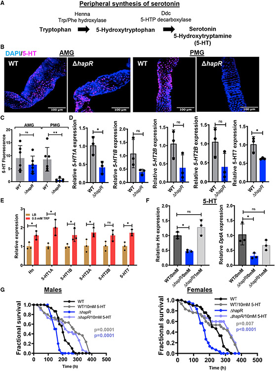
Figure 6. Evidence that V. cholerae-derived tryptophan is converted to serotonin in the host intestine.
(A) Enzymes and intermediates involved in peripheral conversion of tryptophan to serotonin in Drosophila.
(B and C) Representative micrographs (B) and quantification (C) of serotonin immunofluorescence in the AMG and PMG of OreR flies infected with WT V. cholerae or a ΔhapR mutant for 48 h, followed by a 24-h PBS washout. Scale bars, 100 μm. At least six intestines were evaluated. The mean is shown. A Student’s t test was used to calculate significance.
(D and E) qRT-PCR analysis of serotonin receptor expression in the intestines of OreR flies (D) infected with WT V. cholerae or a ΔhapR mutant or (E) supplemented with tryptophan.
(F) qRT-PCR analysis of Hn and DptA expression in the intestines of OreR flies infected with WT or ΔhapR mutant V. cholerae alone or with serotonin (5-HT) supplementation.
(C–F) For qRT-PCR, the mean of biological triplicates is shown. Error bars represent the standard deviation. Significance was calculated using a Student’s t test (C–E) or a one-way ANOVA with Dunnett’s multiple comparisons test (F).
(G) Fractional survival of male and female OreR flies orally infected with WT or ΔhapR mutant V. cholerae alone or with serotonin (5-HT) supplementation. Significance was calculated by log rank analysis.
**p < 0.01, *p < 0.05. See also Table S5.

