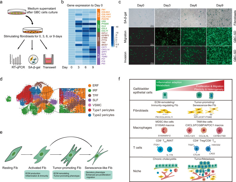
Fig. 8. Dynamic functional states of fibroblasts and dynamics of cellular heterogeneity from inflammation to cancer.
a Workflow displaying in vitro co-culture of human fetal lung fibroblasts (HFL-1) with medium supernatant of GBC-SD cells for 0, 3, 6, and 9 days, respectively. b Heatmap showing expressions dynamics of phenotypic gene signatures in HFL-1 cells when treated with medium supernatant of GBC cells, based on quantitative PCR analysis. c Dynamic illustration of senescence-associated β-galactosidase (SA-β-gal) staining of fibroblasts (first row), transwell cell migration assay of GBC-SD cells co-cultured with stimulated HFL-1 cells (second row), and transwell cell invasion assay of GBC-SD cells co-cultured with stimulated HFL-1 cells (third row) at 0, 3, 6, and 9 days, respectively. Scale bars, 200 μm. d UMAP visualization of the developmental trajectory of mesenchymal cellular subsets inferred by RNA velocity. e Diagram showing functional state transitions of different fibroblasts subtypes. f Schematic illustration overviewing dynamics of cellular heterogeneity from CC to GBC.

