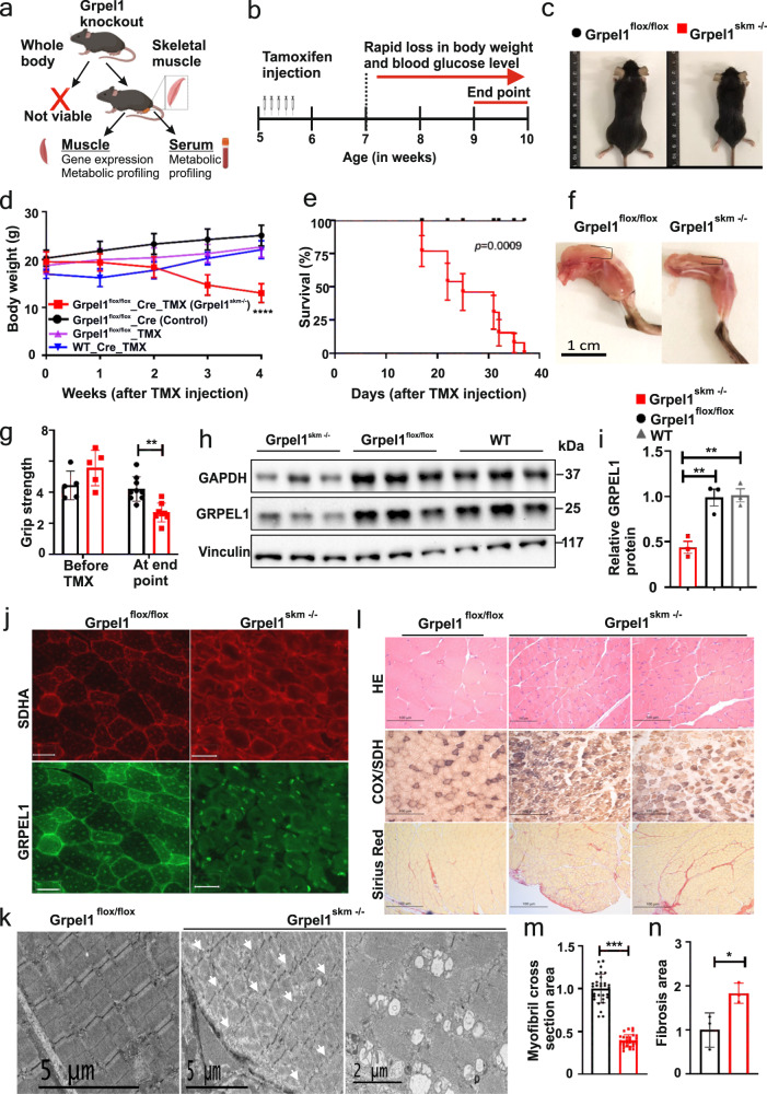
Fig. 1. Inducible loss of mitochondrial co-chaperone Grpel1 in skeletal muscle of mice causes rapid muscular atrophy, retarded growth and shortened lifespan.
a A schematic for the study of whole-body and skeletal muscle-specific knockouts of Grpel1 (Created with BioRender.com). b Timeline of Cre-activation in mice by tamoxifen (TMX) injections. c Representative images of Grpel1skm−/− (right) 4 weeks after tamoxifen injection and the control littermate Grpel1flox/flox (left). d Body weight curve of male mice, starting from TMX injection. Four different groups of mice are shown: Grpel1skm−/− are the inducible skeletal muscle-specific Grpel1 knockout mice, Grpel1flox/flox_Cre are the floxed Cre mice without tamoxifen, Grpel1flox/flox_TMX are the floxed mice without Cre but with tamoxifen, and WT_Cre_TMX are wild type Cre mice with tamoxifen. (n = 10–12 per genotype). Tukey’s multiple comparisons test. ∗∗∗∗p ≤ 0.0001. e Survival curve of Grpel1skm−/− mice analysed with Gehan-Breslow-Wilcoxon test. (n = 13 per genotype). f Representative images of quadriceps of Grpel1skm−/− (right) and control littermate Grpel1flox/flox (left) at sacrification point. g Grip strength of mice immediately before tamoxifen injection and at end point (n = 5–7 per genotype). h Immunoblot showing reduced GRPEL1 level in skeletal muscle of Grpel1skm−/−, in comparison to Grpel1flox/flox and wild type (WT) mice. Vinculin is shown as loading control. i Quantification of GRPEL1 protein level in Grpel1skm−/− muscle from immunoblot against Vinculin (n = 3 per genotype). j Representative immunohistochemistry images of quadriceps femoris (QF) from control and knockout mice with GRPEL1 and SDHA antibodies. Scale bar is 100 µm. k Representative electron microscopic images of QF muscle from control and knockout mice, showing the uneven width of sarcomere and discontinuous Z-line in knockout (left, arrows) and large vacuole structures identified in some fibers (right). l Representative Hematoxylin/Eosin, COX/SDH, and Sirius Red (fibrosis) staining images of QF muscle. m Quantification of cross-sectional area of myofibril in QF muscle (n = 30 representative myofibrils from 3 mice per genotype). n Quantification of fibrotic area of QF muscle (n = 3 mice per genotype).

