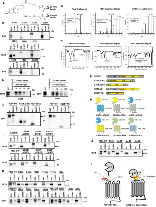
Figure 1.

Cholesterol specifically and reversibly binds to Fzd5, depending primarily on conserved residues at the extracellular linker and loop. A) Demonstration of synthesized 22‐NHC beads and control beads. B) In vitro pulldown assay and WB of Fzd1‐10 by 22‐NHC or control beads. C) Mass‐spectrometry detection of Fzd5‐bound cholesterol (m/z range: 200–600). The cholesterol signature peak at m/z 437.1657 (combined particle of cholesterol, formic acid, and photon) was clearly seen in Fzd5‐associated lipids. Pure cholesterol and Fzd7‐associated lipids are used as positive and negative controls, respectively. D) FT‐IR showing the stretching vibration of ‐OH and ‐CH3/CH2 and the bending vibration of ‐CH3/CH2 (cholesterol signature IR absorption) were clearly seen in Fzd5‐associated lipids. Pure cholesterol and Fzd7‐associated lipids are used as positive and negative controls, respectively. E) 22‐NHC beads pulldown and competition assay of Fzd5 and Smo by cholesterol as a competitor. F) Schematic of various Fzd5 truncation constructs. FL: full‐length; ΔCRD: deletion of CRD; ΔECD: deletion of extracellular domain; ΔC: deletion of the carboxyl terminus; 7TM: transmembrane domain only. G) 22‐NHC beads pulldown assay of Fzd5 truncation constructs depicted in (F). H) Schematic of CRD‐swapped chimeric Fzds. (i.e.: CRD5+ΔCRD7 represents the chimeric protein consisting of Fzd5 CRD and Fzd7 ΔCRD.) I) 22‐NHC beads pulldown assay of the chimeric Fzds depicted in (H). J) 22‐NHC beads pulldown assay of Fzd5 full‐length or truncation of linker segment 1–4 (ΔLS1‐4). K) 22‐NHC beads pulldown assay of Fzd5 WT and point‐mutations of all conserved residues on LS3. L) Working model of cholesterol binding to Fzd5: owing to the steric hindrance, cholesterol could not insert into the hydrophobic grove in the CRD as PA does; the exposed hydrophobic part of cholesterol appears to be covered up by the hydrophobic and aromatic residues in the extracellular linker and loop regions, which form a “cove” with CRD possibly acting as “cap” to wrap around cholesterol. WCL: whole cell lysate.
