
Figure 4.
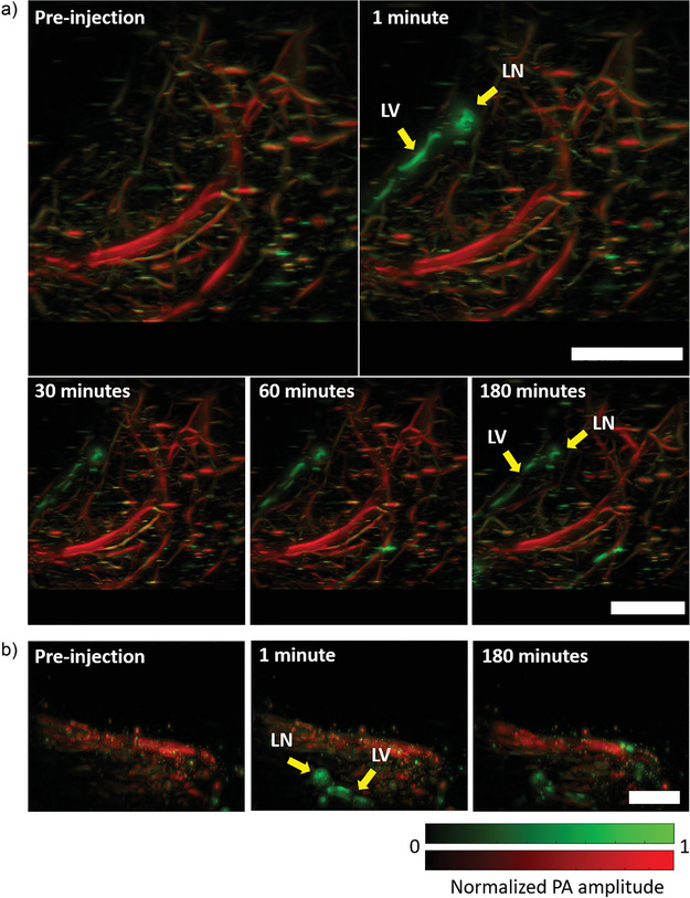
Long‐duration photoacoustic imaging of deep lymphatic vessels in mouse. a) Top‐view images of deep medial lymphatic vessel and lymph node (green), and blood (red) in mouse leg and thigh taken through PACT. b) Side‐view images of deep medial lymphatic vessel (green) and blood (red) in mouse leg and thigh taken through PACT. LV: lymphatic vessel; LN: lymph node; PA: photoacoustic. Scale bars: 0.4 cm.