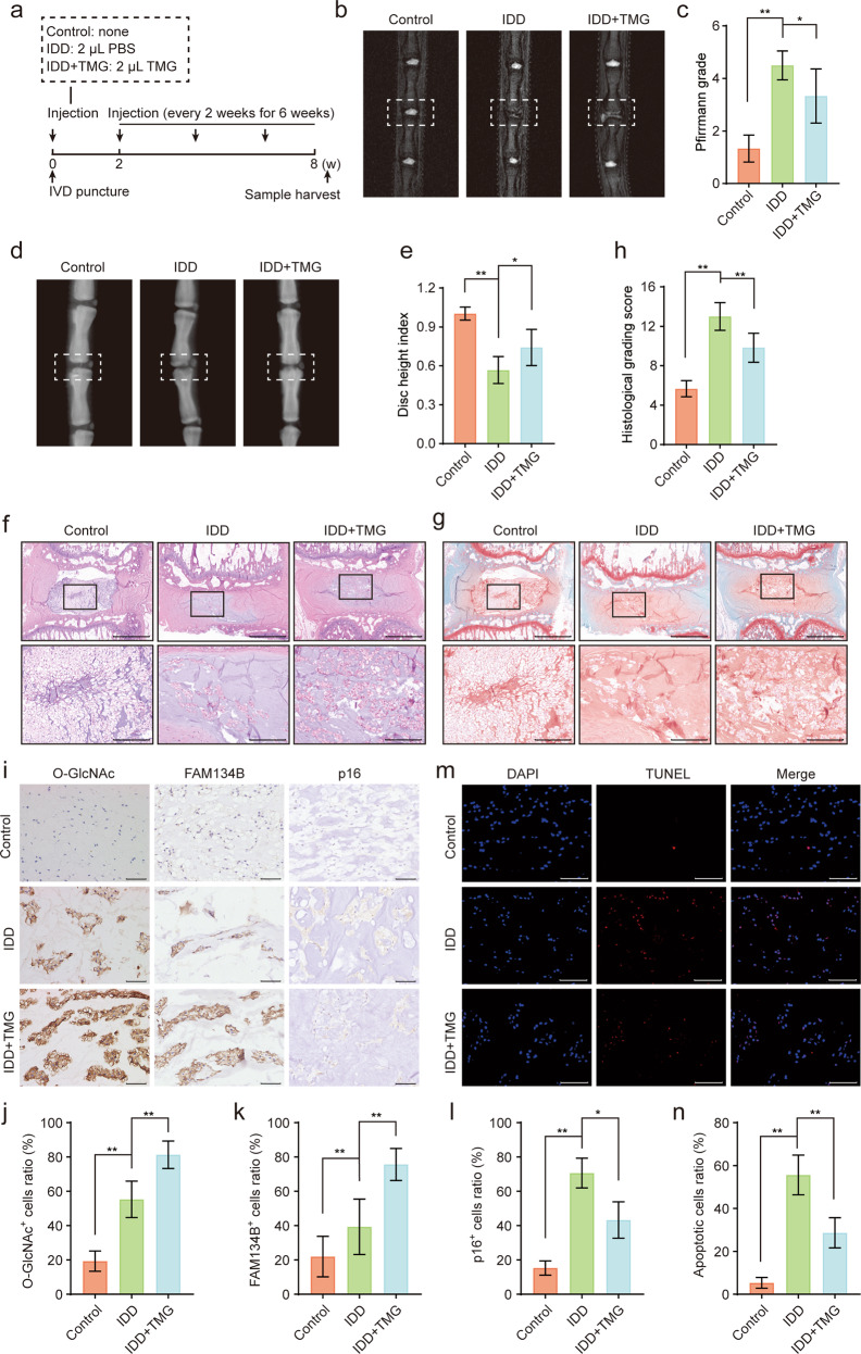
Fig. 6. Administration of TMG partially ameliorates IDD in rats.
a Schematic illustration of the animal model construction and treatments in this study. b, c After 8 weeks of the indicated treatment, the degree of disc degeneration of rat caudal discs was evaluated by T2-weighted MRI and the corresponding Pfirrmann MRI grades were determined. Representative images are shown. d, e Changes in rat caudal disc height were assessed by X-ray imaging. Representative X-ray images were acquired and evaluated on the basis of the DHI. f–h Representative images showing HE and S-O staining of midsagittal sections of rat disc specimens and the corresponding quantitative histologic scores; scale bars: 1 mm and 250 μm. i–l Representative images of O-GlcNAc, FAM134B and p16 immunohistochemical staining of midsagittal sections of rat disc specimens. The immunopositively stained cell to total cell ratio was calculated. Scale bar: 100 μm. m, n Representative images of TUNEL in midsagittal sections of rat disc specimens. Nuclei were stained with DAPI, and positive TUNEL-stained cells were quantified; scale bar: 200 μm. The data are presented as the mean ± SD values. ∗∗P < 0.01, ∗P < 0.05.

