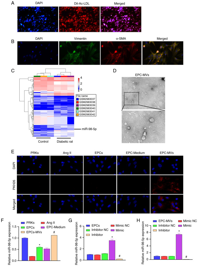
Figure 1.
Recovery of Ang II-induced injury of PRKs and co-culture with EPC-MVs. (A) Confirmation of the isolation of EPCs using Dil-Ac-LDL staining (magnification, x100). (B) Confirmation of the isolation of PRKs through immunofluorescence assay (magnification, x400). (C) The GSE110231 dataset was analyzed using Gene Expression Omnibus to screen miRNAs. This heat map shows differentially expressed miRNAs in diabetic rats compared with healthy rats. Blue bands represent low expression, and red bands represent high expression. (D) Analysis of isolated MVs using transmission electron microscopy (upper image magnification, x15,000; lower image magnification, x40,000). (E) PKH26-labeled EPC-MVs successfully fused with PRKs (magnification, x100). (F) RT-qPCR analysis of the impact of Ang II, EPCs, EPC-Medium and EPC-MVs on the expression of miR-98-5p in PRKs (n=3). *P<0.05 vs. Ang II; #P<0.05 vs. EPC-Medium. (G) RT-qPCR analysis of the impact of miR-98-5p mimic/inhibitor transfection on the expression of miR-98-5p in EPCs (n=3). *P<0.05 vs. mimic NC; #P<0.05 vs. inhibitor NC. (H) RT-qPCR analysis of the impact of miR-98-5p mimic/inhibitor transfection on the expression of miR-98-5p in EPC-MVs (n=3). *P<0.05 vs. mimic NC; #P<0.05 vs. inhibitor NC. Dil-Ac-LDL, Dil complex acetylated low-density lipoprotein; Ang II, Angiotensin II; EPCs, endothelial progenitor cells; PRKs, primary renal kidney cells; MVs, microvesicles; RT-qPCR, reverse transcription-quantitative PCR; NC, negative control; α-SMA, α-smooth muscle actin; miR, microRNA.

