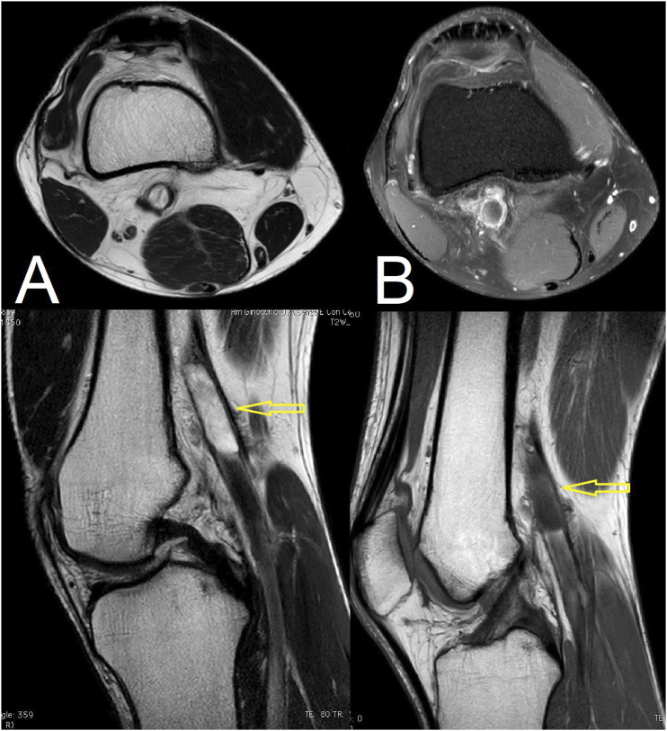
Figure 2.
A: T2-weighted magnetic resonance images. Multi-loculated, high-intensity cystic mass measuring 25 × 30 × 45 mm is located in right popliteal fossa, under: Sagittal view shows popliteal artery surrounded and compressed by cystic mass (white arrow). (B): T1-weighted magnetic resonance images. Multi-loculated, high-intensity cystic mass measuring 25 × 30 × 45 mm is located in right popliteal fossa, under: Sagittal view shows popliteal artery surrounded and compressed by cystic mass (white arrow).

