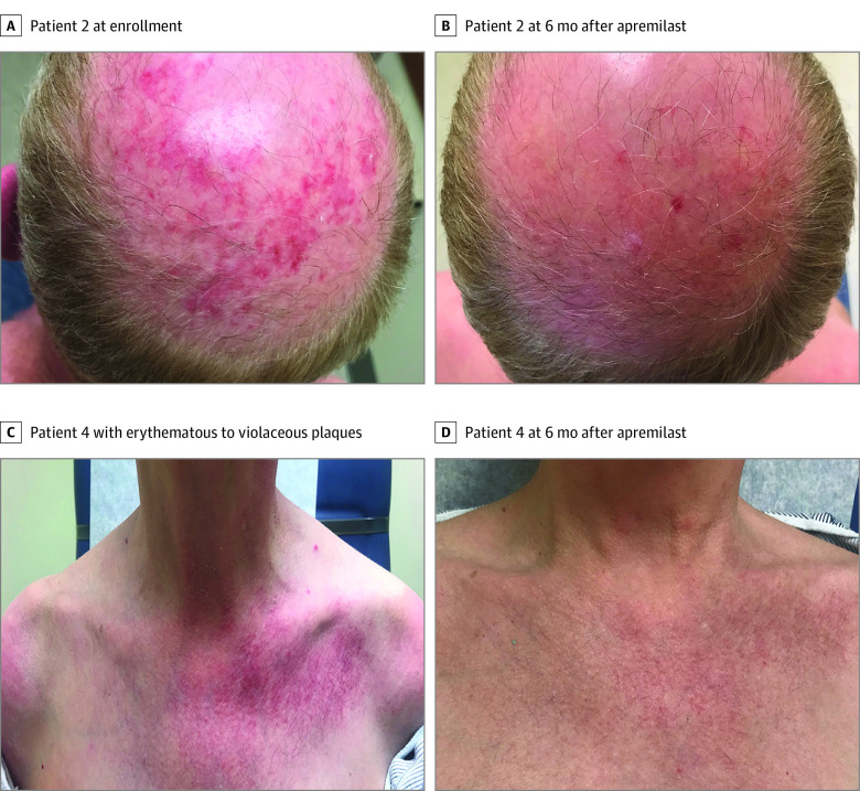
Figure 1. Clinical Photographs.
A, Patient 2, with erythematous plaques with white scale on the scalp at enrollment. B, Patient 2, with resolution of active lesions 6 months after apremilast. C, Patient 4, with erythematous to violaceous plaques with white scale in a characteristic V neck distribution on the chest. D, Patient 4, with resolution of active lesions 6 months after apremilast with remaining faint telangiectasia.

