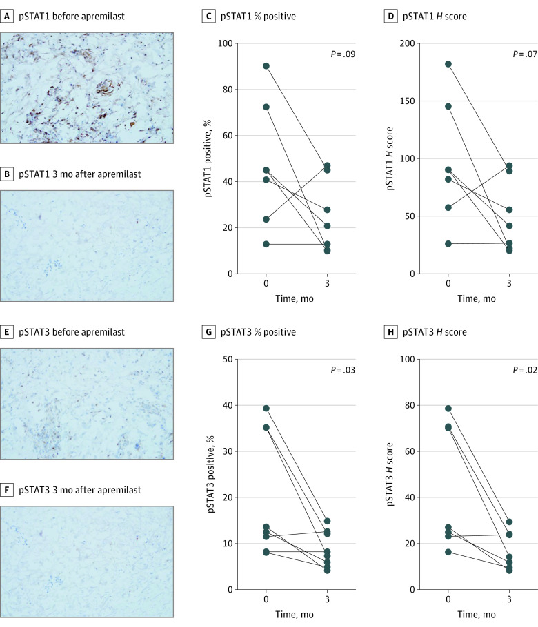
Figure 4. Association of Apremilast With Protein Phosphorylation on Immunohistochemistry.
A-B, Decrease in phosphorylated signal transducers and activators of transcription 1 (pSTAT1) immunostaining on a frozen section of patient 2 skin biopsies before and 3 months after apremilast (original magnification ×200). C-D, Paired analysis change in pSTAT1 percent positive cells and H score at baseline and 3 months after apremilast. E-F, Decrease in pSTAT3 immunostaining on frozen section from patient 2 skin biopsies before and 3 months after apremilast (original magnification ×200). G-H, Paired analysis change in pSTAT3 percent positive cells and H score at baseline and 3 months after apremilast.

