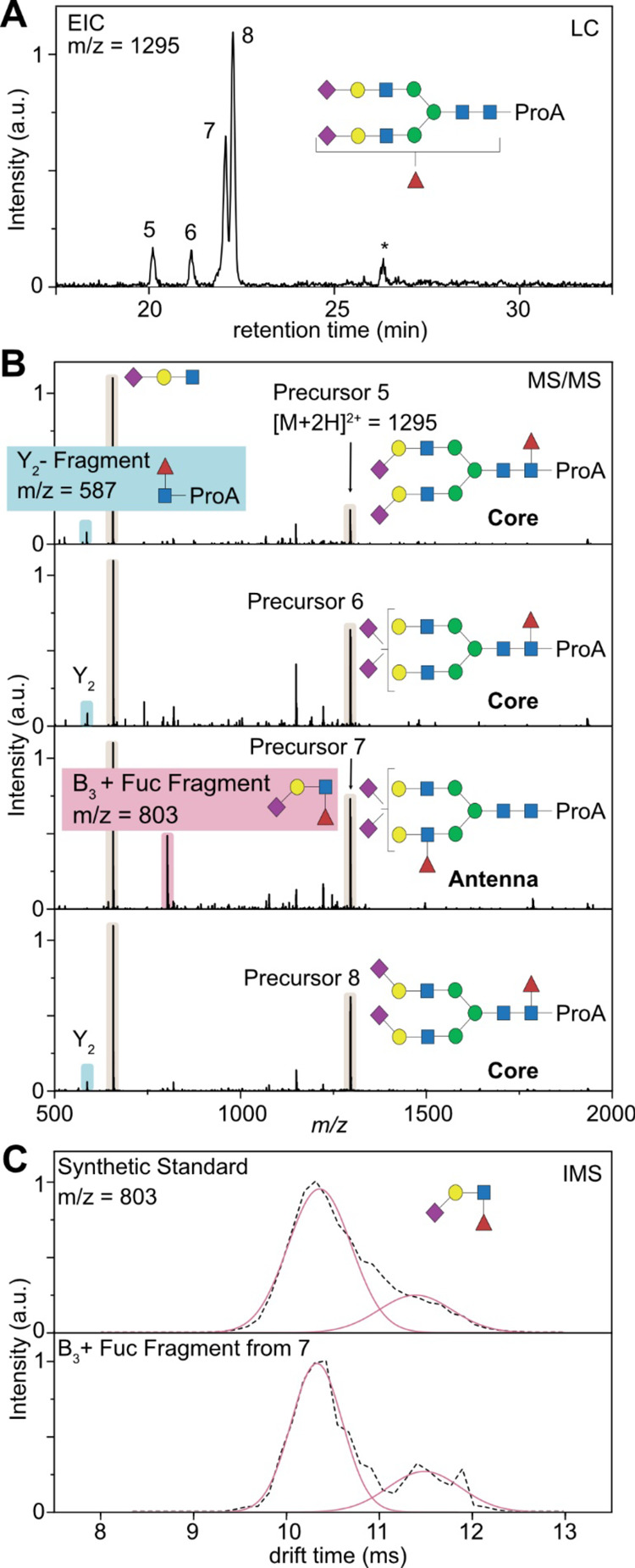
Figure 5.

Determination of the fucosylation pattern based on HILIC-IM-MS. (A) Extracted ion chromatogram (EIC) of a doubly sialylated biantennary glycan with one fucose attached (m/z = 1295). Minor peaks marked with an asterisk are fragment ions generated from larger glycans. (B) MS/MS spectra of the precursor ions 5–8, which are almost identical and show the dominant B3 trisaccharide fragment. One major difference stems from either Y2 fragmentation (highlighted in blue) or B3 fragmentation (highlighted in red). (C) Mobilograms of the B3 + fucose fragment generated from precursor ion 7. Comparison with a synthetic standard (3′-Sialyl-Lewis-X) allows us to identify the fucose isoforms and confirm the native state of the fucosylation.
