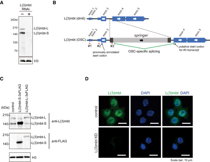
Figure EV1. Two isoforms of L(3)mbt, L(3)mbt‐L, and L(3)mbt‐S, in OSCs.

- Western blotting using an anti‐L(3)mbt antibody that we raised in this study detected L(3)mbt as a doublet in the OSC nuclear lysates. The doublet disappeared upon L(3)mbt RNAi. Histone H3 (H3) served as a loading control.
- The exon‐intron structures of the l(3)mbt gene in dm6 (provided by UCSC) and in OSCs (Sienski et al, 2012). Note that the l(3)mbt gene in OSCs has an LTR‐type transposon, springer. RACE detected three different 5′ ends of l(3)mbt mRNA (#1, #2, and #3).
- Top: Western blotting using the anti‐L(3)mbt antibody shows that L(3)mbt‐L‐3xFLAG and L(3)mbt‐S‐3xFLAG exogenously expressed in OSCs co‐migrate with endogenous L(3)mbt‐L and L(3)mbt‐S (control), respectively. Middle: Western blotting was performed using anti‐FLAG antibody. Bottom: Histone H3 (H3) was detected as a loading control.
- Immunofluorescence analysis using the anti‐L(3)mbt antibody detected L(3)mbt (green) mostly in the OSC nuclei. The L(3)mbt signals disappeared upon L(3)mbt RNAi (KD). DAPI (blue) indicates the nuclei. Scale bar: 10 μm.
Source data are available online for this figure.
