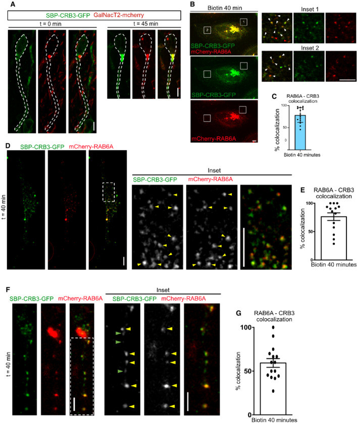
Figure EV3. CRB3 exits the Golgi within RAB6+ vesicles.

-
ASBP‐CRB3‐GFP and GalNacT2‐mCherry expression in aRG cells before and 45 min after addition of biotin. SBP‐CRB3‐GFP relocates from a diffuse perinuclear localization to the Golgi. Scale bar = 5 μm.
-
BSBP‐CRB3‐GFP and mCherry‐RAB6A localization in HeLa cells before and 40 min after addition of biotin. Scale bar = 5 μm. White arrowheads: colocalizing foci.
-
CQuantification of SBP‐CRB3‐GFP and mCherry‐RAB6A colocalization away from the Golgi apparatus 40 min after biotin addition. N = 16 cells from three independent experiments.
-
DSBP‐CRB3‐GFP and mCherry‐RAB6A localization in dissociated aRG cells cultivated in vitro, 40 min after addition of biotin. Scale bar = 5 μm. Yellow arrowheads: colocalizing foci.
-
EQuantification of SBP‐CRB3‐GFP and mCherry‐RAB6A colocalization away from the Golgi apparatus 40 min after biotin addition. N = 14 cells from three independent experiments.
-
FSBP‐CRB3‐GFP and mCherry‐RAB6A localization in aRG cells cultivated within brain slices, 40 min after addition of biotin. Scale bar = 5 μm. Yellow arrowheads: colocalizing foci.
-
GQuantification of SBP‐CRB3‐GFP and mCherry‐RAB6A colocalization away from the Golgi apparatus 40 min after biotin addition. N = 15 cells from three independent experiments.
Data information: All error bars indicate SD.
