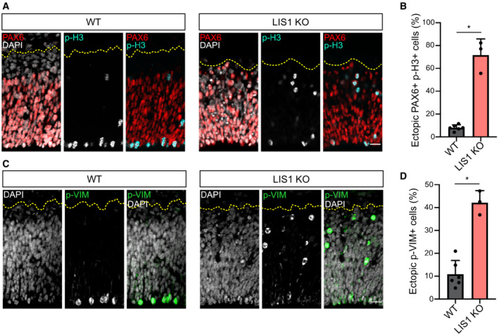
Figure 5. LIS1 knockout leads to ectopically dividing progenitors.

-
APAX6 and phospho‐Histone 3 (p‐H3) staining in WT and LIS1 KO E12.5 brains. Cortices were subdivided into five bins of equal size along the radial axis. Scale bar = 25 μm.
-
BPercentage of PH3+/PAX6+ cells located above the ventricular surface of WT and LIS1 KO E12.5 brains. WT: 1192 cells from N = 6 brains. LIS1 KO: 589 cells from N = 3 brains.
-
CPhospho‐Vimentin (p‐VIM) staining in WT and LIS1 KO E12.5 brains. Scale bar = 25 μm.
-
DPercentage p‐VIM+ cells dividing ectopically, away from the ventricular surface of WT and LIS1 KO E12.5 brains. WT: 1056 cells from N = 6 brains. LIS1 KO: 879 cells from N = 3 brains.
Data information: (B, D) Mann–Whitney U test, *P ≤ 0.05. All error bars indicate SD.
