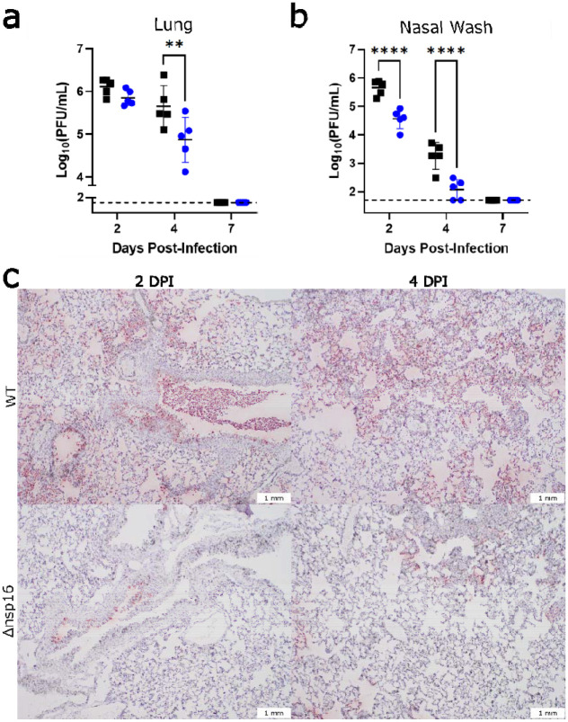
Figure 4. dNSP16 replication is reduced in vivo.
(a, b) Comparison of viral titers from (a) right cranial lung lobes or (b) nasal washes from WT- (black) or dNSP16-infected (blue) hamsters sacrificed at the indicated day. **p<0.01, ****p<0.001: results of two-way ANOVA with Tukey’s multiple comparison test (α = 0.05). Values from individual hamsters are plotted (symbols) as well as means (black bars). Error bars denote standard deviation. Dotted lines represent limits of detection. PFU = plaque-forming units. (c) SARS-CoV-2 nucleocapsid staining (brown) of representative 5 μm-thick sections taken from left lung lobes.

