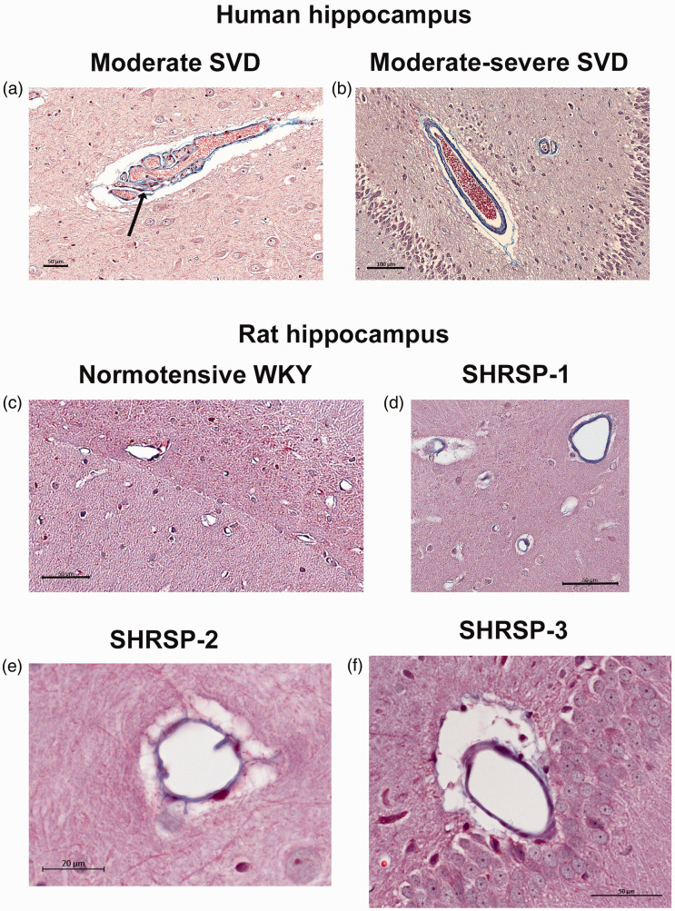
Figure 1.
Human post-mortem hippocampal samples compared to SHRSP rat brain (a) Post-mortem Masson’s trichrome stained hippocampal section from a subject diagnosed with moderate cerebral small vessel disease (SVD). This diagnosis was supported by evidence of fibrosis/hyalinosis of the small arteries (blue-color in the vessel wall). The tortuous arteriole with collagenous fibrosis is surrounded by an enlarged PVS. A Charcot-Bouchard microaneurysms can also be noted (arrow). (b) Post-mortem Masson’s trichrome stained hippocampal section from a subject diagnosed with moderate-to-severe case where more extensive collagenous fibrosis is noted at the level of the arteries and arterioles. (c) Trichome-stained section from a normotensive WKY rats at the level of the hippocampal sulcus demonstrating minimal collagenous fibrosis associated with arterioles and (d–f) Masson’s trichrome stained hippocampal sections from three different SHRSP rat showing more extensive collagenous fibrosis and a dilated PVS consistent with ‘moderate’-like SVD. Scale bars: a, c, d, f: 50 µm, b: 100 µm and e: 20 µm.

