
Figure 1. The single-cell immunobiology of the maternal-fetal interface in early pregnancy.
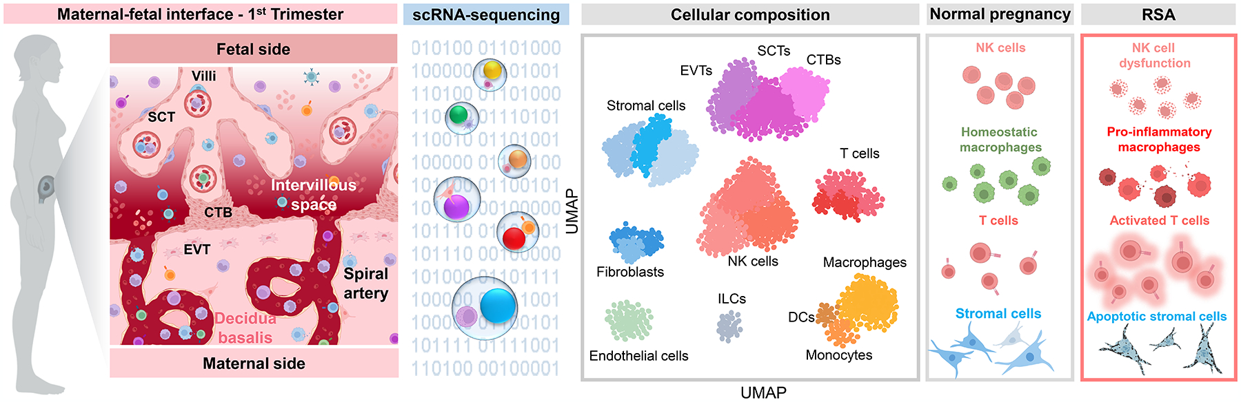
The first-trimester maternal-fetal interface is marked by placental growth, trophoblast invasion and angiogenesis, and the establishment of maternal-fetal dialogue to promote homeostasis for the remainder of pregnancy. During this period, the placental compartment primarily comprises trophoblast cell types, fibroblasts, stromal cells, and Hofbauer cells (fetal macrophages), as represented in the UMAP plot based on data reported by Vento-Tormo et al. 2018. By contrast, the decidua displays more heterogeneous cellularity, characterized by major populations of decidual stromal cells, NK cells, macrophages, and endothelial cells. Other leukocyte subsets are also represented in the early decidua; namely, T cells, innate lymphoid cells, and dendritic cells. Single-cell investigations of recurrent spontaneous abortion (RSA) have suggested that this disease includes the altered composition of decidual NK cells and generalized T-cell infiltration and activation, together with the acquisition of a pro-inflammatory phenotype by NK cells and macrophages. Indeed, it was proposed that a shift away from macrophage-NK cell interactions toward macrophage-T cell interactions may contribute to RSA. The decidual stromal compartment may also be affected, as enrichment of cellular processes related to apoptosis and senescence implicates impaired stromal cell differentiation in RSA pathophysiology. Abbreviations used: SCT, syncytiotrophoblast; CTB, cytotrophoblast; EVT, extravillous trophoblast; NK cell, natural killer cell; DC, dendritic cell; ILC, innate lymphoid cell; UMAP, uniform manifold approximation and projection. Figure created using BioRender.