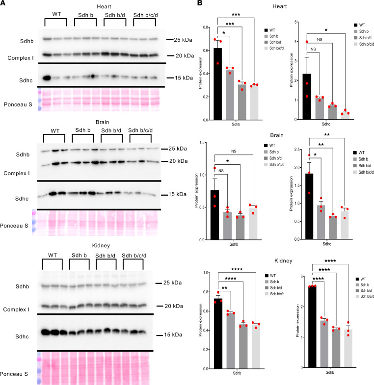
Figure 1. Protein levels of SDHs decrease in Sdh hKO mice.
(A) Western blots of Sdhb, Sdhc, and complex I protein NADH:ubiquinone oxidoreductase subunit B8 (NDUFB8) from heart, kidney, and brain tissues from WT (n = 3), Sdhb (n = 3), Sdhb/d (n = 3), and Sdhb/c/d (n = 3) mice. Ponceau S staining was used as a loading control. The experiment is conducted once. (B) Quantification of bands’ intensity as a measurement of protein levels from heart, kidney, and brain tissues from WT (n = 3), Sdhb (n = 3), Sdhb/d (n = 3), and Sdhb/c/d (n = 3) mice. Sdhb and Sdhc were normalized to complex I NDUFB8. P values are calculated by ordinary 1-way ANOVA, (*P < 0.05, **P < 0.01, ***P < 0.001, ****P < 0.0001). Data represent mean ± SEM.

