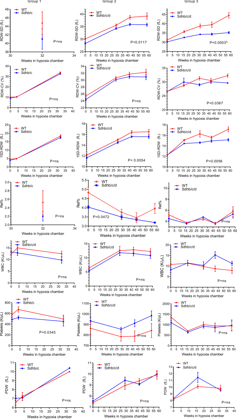
Figure 4. RDW-SD, RDW-CV, 1SD-RDW, reticulocyte percentage, white blood cells, platelets, and platelet distribution width in Sdh hKO (Sdhb/c in group 1 and Sdhb/c/d in groups 2 and 3) and WT control mice under chronic hypoxia.
Measures of RBC size variation including RDW-SD, RDW-CV and 1SD-RDW, reticulocyte percentage (Ret%), white blood cells (WBCs), platelets, and platelet distribution width (PDW) are shown. Each time point contains 3–5 male mice and shows mean and SEM. P values are calculated by 2-way ANOVA using time and genotype as independent variables. AP value remains significant (less than 0.05) after adjustment for multiple comparisons by Holm-Šídák method (α: 0.05). The missing parameters in group 1 (RDW-SD, Ret%) were not available in earlier CBC outputs. The missing parameter IRF in groups 1 and 2 was not available in earlier CBC outputs.

