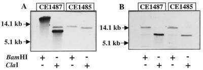
FIG. 5.
Southern blot analysis of chromosomal DNA of CE1485 and CE1487. Blots were hybridized with the pitB probe (A) and probe 3 (B) (see Fig. 4). DNA was digested with BamHI or ClaI, and fragments were separated by electrophoresis on a 0.8% agarose gel. The DNA was transferred from the gel to Hybond-N+ membranes (Amersham) with a vacuum blotter (Bio-Rad model 785). After transfer, the filter was washed in 2× SSC (1× SSC is 0.15 M NaCl plus 0.015 M sodium citrate [pH 7.0]) for 5 min, and DNA was cross-linked by UV irradiation for 2 min. Labeling of the probes, hybridization, and detection were done with digoxigenin labeling and detection kits (Boehringer Mannheim). Hybridization and stringency washes were carried out at 68°C. The positions of molecular size standard DNA fragments are indicated in kilobases.

