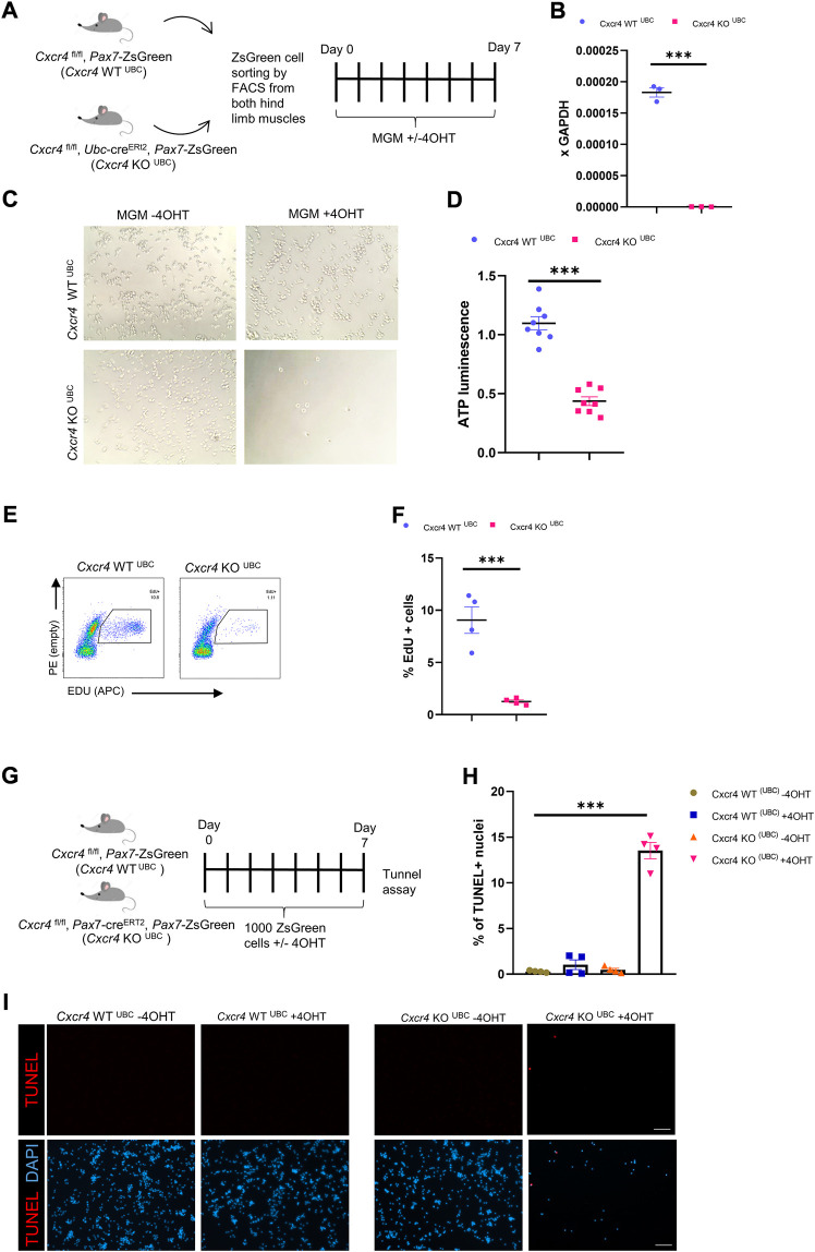
FIGURE 4.
CXCR4 regulates early proliferation of satellite cells. (A) Schematic for Tamoxifen treatment and time of analysis. (B) RTqPCR for expression of Cxcr4 in myoblasts from Cxcr4FL/FL; Ubc-creERT2; Pax7-ZsGreen mice (KOUBC) (n = 3) or Cxcr4 FL/FL; Ubc-creERT2; Pax7-ZsGreen controls lacking cre (WTUBC) (n = 3), treated with 4OHT. (C) Representative bright field images for myoblasts from Cxcr4-KOUBC or WTUBC +/- 4OHT. (D) Cell growth assay measuring ATP luminescence in myoblasts from Cxcr4-KOUBC (n = 3) or WTUBC (n = 3) +/- 4OHT. Results are normalized to untreated. (E) Representative FACS plots for EdU staining of myoblasts 7 days in culture from Cxcr4-WTUBC +4OHT (n = 3) and Cxcr4 KOUBC +4OHT (n = 3). (F) Frequency of EdU + cells for replicates of the cells shown in E. (G) Schematic for the in vitro analysis of apoptosis. (H) Frequency of TUNEL + nuclei in Cxcr4-WTUBC and KOUBC cultured satellite cells ± 4OHT. (I) Representative immunofluorescence staining for TUNEL in Cxcr4-WTUBC and Cxcr4-KOUBC +/- 4OHT scale bar = 100 µm. Note also the low cell number in KOUBC + 4OHT. Data presented as mean ± SEM, ***p < 0.001 by t-test and two-way ANOVA.

